The Opponents
Why do colors have opposites? Why would it be impossible to describe a color as greenish red or yellowish blue? In this part, I’ll explain more about the perception of different colors, and I’ll introduce you to the next level of processing:
The Opponent Process Theory
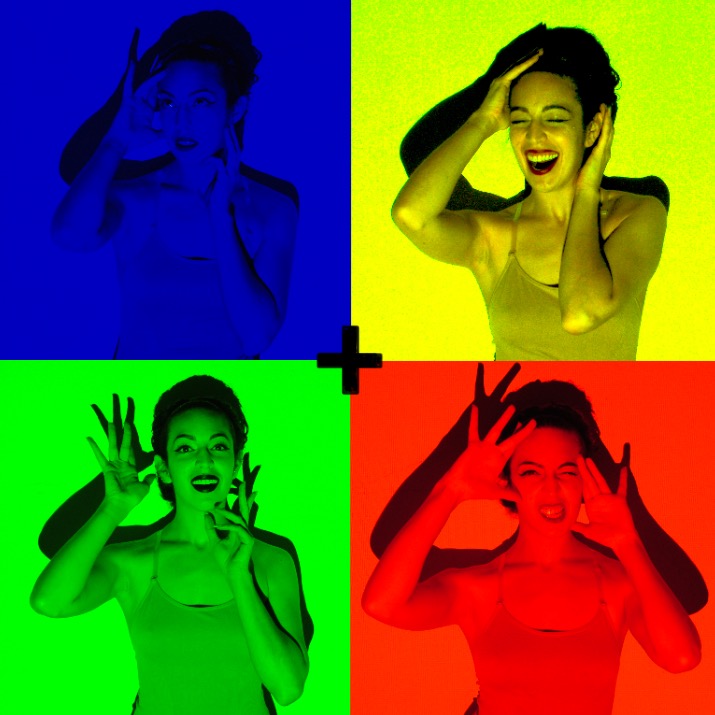
Stare at the black cross in the picture on the left for 30-60 secs. Now look over at the black cross on the left. Any fun effects? (Click on the image to enlarge it, makes for a better effect.)
We last left our conical heroes, the S, M, and L cones, at the retina with the introductory knowledge of how we see the world as a Trichromat. But these are just three colors, how then do we differentiate over a million? Let us continue on this colorful saga. Thus far we have been discussing what happens in the first level of color processing, which involves the rods and cones located in the retina, which is explained by the Trichromatic Color Theory.
“Hmm… could you refresh my memory?”
The retina is a thin layer of tissue lining the inside of your eye. Distributed throughout this tissue are rods and cones, the main cells that detect light in the eye. In most humans, there are three types of cones. There are short wave (400-500nm) S cones, medium wave (450-630nm) M cones, and long wave (500-700nm) L cones. Sometimes the S, M, and L cones are referred to as the Blue, Green, and Red cones, respectively, though they do not directly correspond to just blue, green, and red colors. Cones are activated by wavelengths of varying frequencies, even those that are not in their own maximal range. In other words, a green cone may still be stimulated slightly by a non-green color.
“How do you sort the whole colorful mess out then?”
Beyond the paper thin retinal sheet of rods and cones are another type of cells known as ganglion cells. They are the next operators in the game of telephone from your eyes to your conscious mind. At this level, the Opponent Process Theory arises. Throughout most of the retina most ganglion cells receive information from more than one cone. It is the ganglion cell that has the task of summarizing the information from the multiple cones within a receptive field.
Receptive fields can be pictured as a bull’s-eye with a center and a surrounding area. The center will contain one type of cone, and the surround will be its “opponent”. Depending on the light hitting the receptive field, the center can be activated, or “on”, and the surrounding is inhibited, or “off”. The reverse situation is possible as well, with the center being “off” and the surround being “on”. Color vision at this level involves the comparison of the proportion of nerve impulses from each cone type. To say it another way, the visual system is interested in the differences in cone responses as opposed to each individual cone’s response. There are two chromatic opponent processing pairs: red-green and blue-yellow. Ever notice you never see a reddish-green or a bluish-yellow? It is because the two opponent colors cannot be perceived simultaneously.
“But why not?”
Notice how red and green cones have overlapping responsiveness. A stimulus may activate both green and red cones, but it is the proportions that give rise to discrimination. Let’s assume the stimulus is red. In a red center, green surrounding receptive field, the center, which is responsive to red is “on” and the surrounding is “off”. Alternatively, in a green-red receptive field, the center will be off and the surrounding red will be on. Both red and green cones will be activated but when they make it to the next level of processing, the summation will be in favor of red because in most cases the red center and red surround prevailed. So, rather than receive multiple and mixed messages, the cortex gets the summarized message of, “It’s red, boss.”
“That’s all well and good, but what about this yellow?!”
Ah, yes. Yellow. Let’s take a look at yellow… The wavelength of yellow falls between the maximal green (M cones) and red (L cones) wavelengths. So which cone type is activated? If you guessed both, you are correct! There is also input from the blue-yellow receptive field channels. Because the wavelength is around 570 nm, the blue, S cones are not activated (they prefer below 500nm) which renders the yellow channel dominant. It is the combination of red and green activation and lack of blue activation (along with some additional processing in higher order cortical areas), that gives rise to the perception of yellow.
In addition to giving rise to the perception of a million colors or so, the opponent process mechanism allows for some rather entertaining optical illusion tricks. If you stare at an image of one color such as green then look away to a white wall, you may notice a reddish/magenta afterimage. The green channel is fatigued by overstimulation and loses sensitivity because the green cones exhaust their photopigment supply. This allows for red to be temporarily dominate, creating the afterimage. Scattered through out this article are various afterimage examples to pit your opponent pairs against one another. I do hope you enjoy them.
Until next time my Dichromatic, Trichromatic (perhaps even Tertachromatic?!) friends!
Published: United Academics (Feb. 23, 2015)